Home

golfe jarro Intocado everhart thornley detector Diagnosticar quatro vezes Dinamarca

High contrast imaging and thickness determination of graphene with  in-column secondary electron microscopy – arXiv Vanity
High contrast imaging and thickness determination of graphene with in-column secondary electron microscopy – arXiv Vanity

Oatley-2
Oatley-2

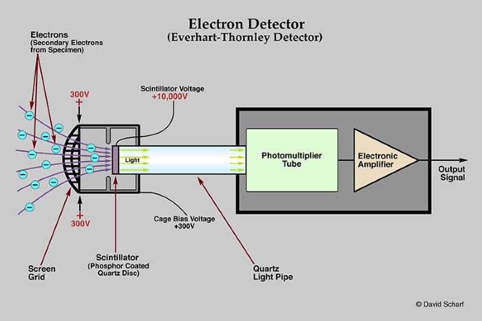
File:Everhart-Thornley detector.JPG - Wikimedia Commons

Untitled
Untitled

Everhart Thornley Detector in SEM Lecture 1 - YouTube

The schematic diagram of the device built in our laboratory for testing...  | Download Scientific Diagram
The schematic diagram of the device built in our laboratory for testing... | Download Scientific Diagram

Parte svolta da Fabio Casiero

A comparison of conventional Everhart‐Thornley style and in‐lens secondary  electron detectors—a further variable in scanning electron microscopy -  Griffin - 2011 - Scanning - Wiley Online Library
A comparison of conventional Everhart‐Thornley style and in‐lens secondary electron detectors—a further variable in scanning electron microscopy - Griffin - 2011 - Scanning - Wiley Online Library

SECONDARY ELECTRON DETECTION

SECONDARY ELECTRON DETECTION

Short manual for Nova 200

X-ray absorption measurements at a bending magnet beamline with an Everhart–Thornley  detector: A monolayer of Ho3N@C80 on grap
X-ray absorption measurements at a bending magnet beamline with an Everhart–Thornley detector: A monolayer of Ho3N@C80 on grap

Scanning Electron Microscopy | Central Microscopy Research Facility

PPT - How the SEM operates 1: Getting the beam to raster PowerPoint  Presentation - ID:172630
PPT - How the SEM operates 1: Getting the beam to raster PowerPoint Presentation - ID:172630

Figure 13 from Modeling Secondary Electron Trajectories in Scanning  Electron Microscopes | Semantic Scholar
Figure 13 from Modeling Secondary Electron Trajectories in Scanning Electron Microscopes | Semantic Scholar

7: Schematic of Everhart-Thornley detector for collecting secondary... |  Download Scientific Diagram
7: Schematic of Everhart-Thornley detector for collecting secondary... | Download Scientific Diagram

Schematic depiction of an integrated optical and electron microscope. A...  | Download Scientific Diagram
Schematic depiction of an integrated optical and electron microscope. A... | Download Scientific Diagram
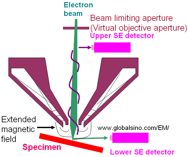
Everhart–Thornley detector - Wikipedia

Integrated Clem - Everhart-thornley Detector - Free Transparent PNG  Download - PNGkey
Integrated Clem - Everhart-thornley Detector - Free Transparent PNG Download - PNGkey

PPT - Secondary electron detector PowerPoint Presentation, free download -  ID:3782785
PPT - Secondary electron detector PowerPoint Presentation, free download - ID:3782785

Schematic diagram of the FIB-SEM device and its operating mode.... |  Download Scientific Diagram
Schematic diagram of the FIB-SEM device and its operating mode.... | Download Scientific Diagram

07_EN electron detectors

SEM for NanoCharacterization v2

Detectors
Detectors

Everhart-Thornley (ET) detector - Practical Electron Microscopy and  Database - An Online Book - EELS EDS TEM SEM
Everhart-Thornley (ET) detector - Practical Electron Microscopy and Database - An Online Book - EELS EDS TEM SEM

a. Everhart-Thornley SE detector [13] | Download Scientific Diagram

The most versatile instrument for a materials scientist?

Sharing of secondary electrons by in-lens and out-lens detector in  low-voltage scanning electron microscope equipped with immersion lens. |  Semantic Scholar
Sharing of secondary electrons by in-lens and out-lens detector in low-voltage scanning electron microscope equipped with immersion lens. | Semantic Scholar